Home

Победен полярен дванадесет ζώνες giemsa регулируем Мелбърн едва

Lymphocyte nuclei of nodal marginal zone lymphoma mimicking granulocytic  morphology with Pelger–Huët-like features - Pathology
Lymphocyte nuclei of nodal marginal zone lymphoma mimicking granulocytic morphology with Pelger–Huët-like features - Pathology

The immune cells by Giemsa stain (A, B) and toluidine blue (C, D). (A,... |  Download Scientific Diagram
The immune cells by Giemsa stain (A, B) and toluidine blue (C, D). (A,... | Download Scientific Diagram

Figure 5 | A combined banding method that allows the reliable  identification of chromosomes as well as differentiation of AT- and GC-rich  heterochromatin | SpringerLink
Figure 5 | A combined banding method that allows the reliable identification of chromosomes as well as differentiation of AT- and GC-rich heterochromatin | SpringerLink

PLOS Medicine: Evaluation of splenic accumulation and colocalization of  immature reticulocytes and Plasmodium vivax in asymptomatic malaria: A  prospective human splenectomy study
PLOS Medicine: Evaluation of splenic accumulation and colocalization of immature reticulocytes and Plasmodium vivax in asymptomatic malaria: A prospective human splenectomy study

View Image
View Image

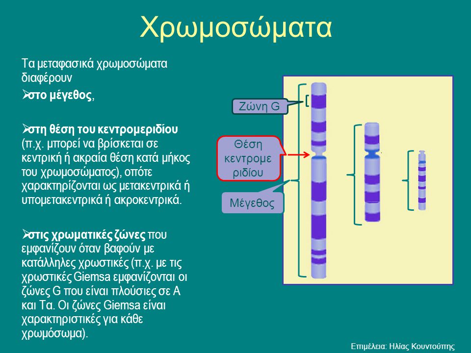
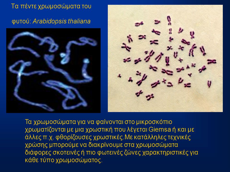
Η οργάνωση του γενετικού υλικού - ppt κατέβασμα

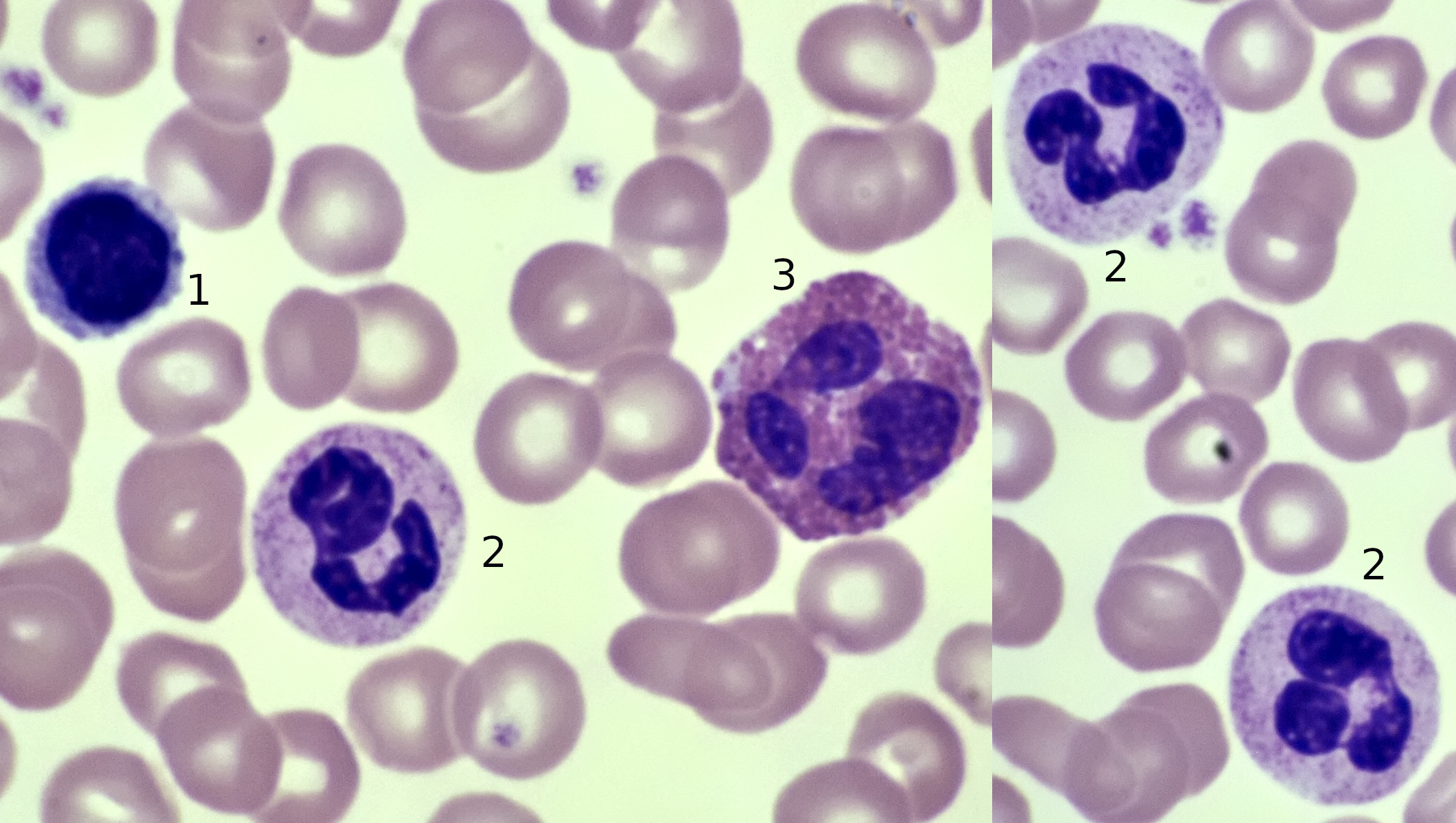
Blood | Microscopy of Nature

Η οργάνωση του γενετικού υλικού - ppt κατέβασμα

Bone marrow aspiration smear (May-Giemsa staining, ×1,000). Note: The... |  Download Scientific Diagram
Bone marrow aspiration smear (May-Giemsa staining, ×1,000). Note: The... | Download Scientific Diagram

Bone marrow histology in marginal zone B-cell lymphomas: correlation with  clinical parameters and flow cytometry in 120 patients - Annals of Oncology
Bone marrow histology in marginal zone B-cell lymphomas: correlation with clinical parameters and flow cytometry in 120 patients - Annals of Oncology

Absence of elastic fibers in the central zone (Orcein- Giemsa staining,...  | Download Scientific Diagram
Absence of elastic fibers in the central zone (Orcein- Giemsa staining,... | Download Scientific Diagram
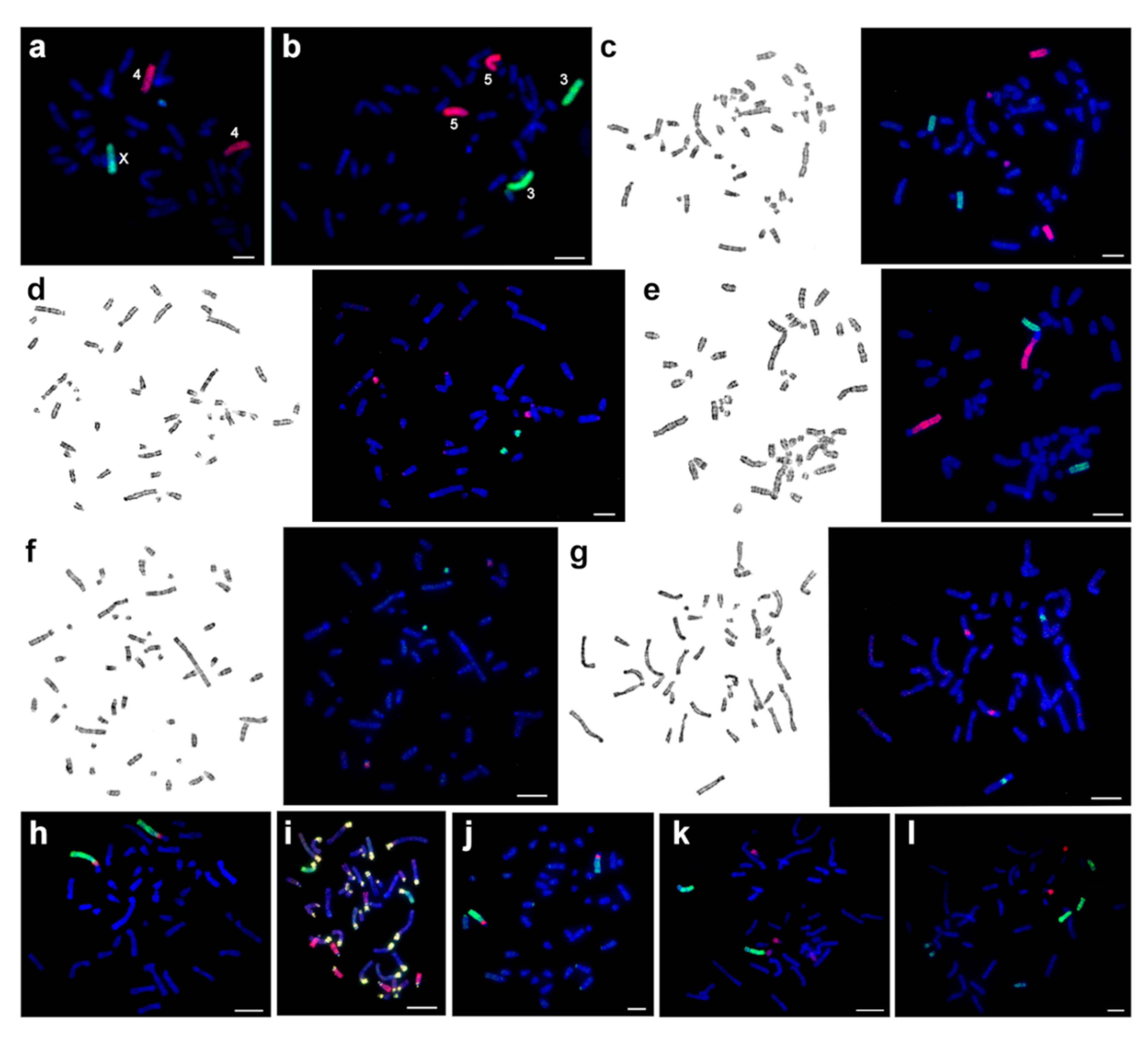
Genes | Free Full-Text | New Data on Comparative Cytogenetics of the  Mouse-Like Hamsters (Calomyscus Thomas, 1905) from Iran and Turkmenistan |  HTML
Genes | Free Full-Text | New Data on Comparative Cytogenetics of the Mouse-Like Hamsters (Calomyscus Thomas, 1905) from Iran and Turkmenistan | HTML
Giemsa stain encapsulated Cryptococcus neoformans: Introduction

Peripheral blood smear stains with May-Grünwald Giemsa: (a) Low... |  Download Scientific Diagram
Peripheral blood smear stains with May-Grünwald Giemsa: (a) Low... | Download Scientific Diagram

Restoring production of hippocampal neurons in old age | Nature Neuroscience

ΧΡΩΜΟΣΩΜΑΤΑ Τα χρωμοσώματα είναι η μορφή που παίρνει η χρωματίνη - ppt  κατέβασμα
ΧΡΩΜΟΣΩΜΑΤΑ Τα χρωμοσώματα είναι η μορφή που παίρνει η χρωματίνη - ppt κατέβασμα

Candida glabrata in Giemsa stain of sputum: Introduction, Pathogenecity

Figure 3 | Diagnostic Workup of Small B Cell Lymphomas: A Laboratory  Perspective
Figure 3 | Diagnostic Workup of Small B Cell Lymphomas: A Laboratory Perspective

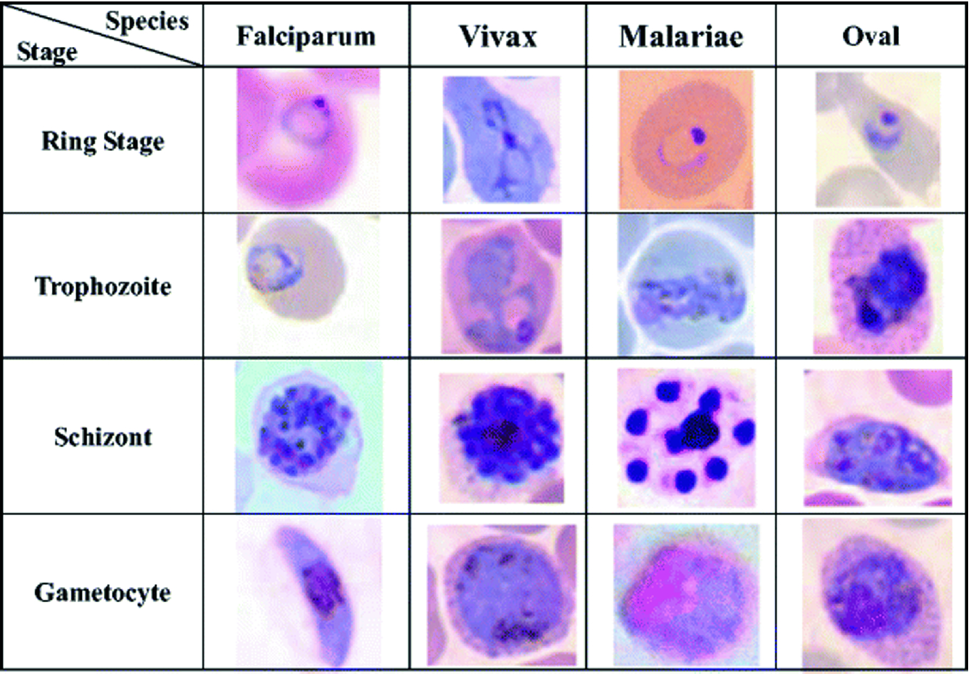
Malaria Detection on Giemsa-Stained Blood Smears Using Deep Learning and  Feature Extraction | SpringerLink
Malaria Detection on Giemsa-Stained Blood Smears Using Deep Learning and Feature Extraction | SpringerLink
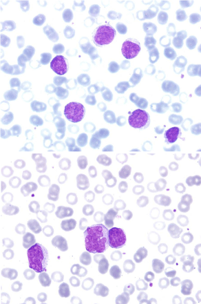
A Case of Splenic Marginal Zone Lymphoma with Mismatched Morphology and  Phenotype, Karyotype and Clinical Course
A Case of Splenic Marginal Zone Lymphoma with Mismatched Morphology and Phenotype, Karyotype and Clinical Course
Lymph node biopsy consistent with marginal zone lymphoma (40×,... |  Download Scientific Diagram
Lymph node biopsy consistent with marginal zone lymphoma (40×,... | Download Scientific Diagram

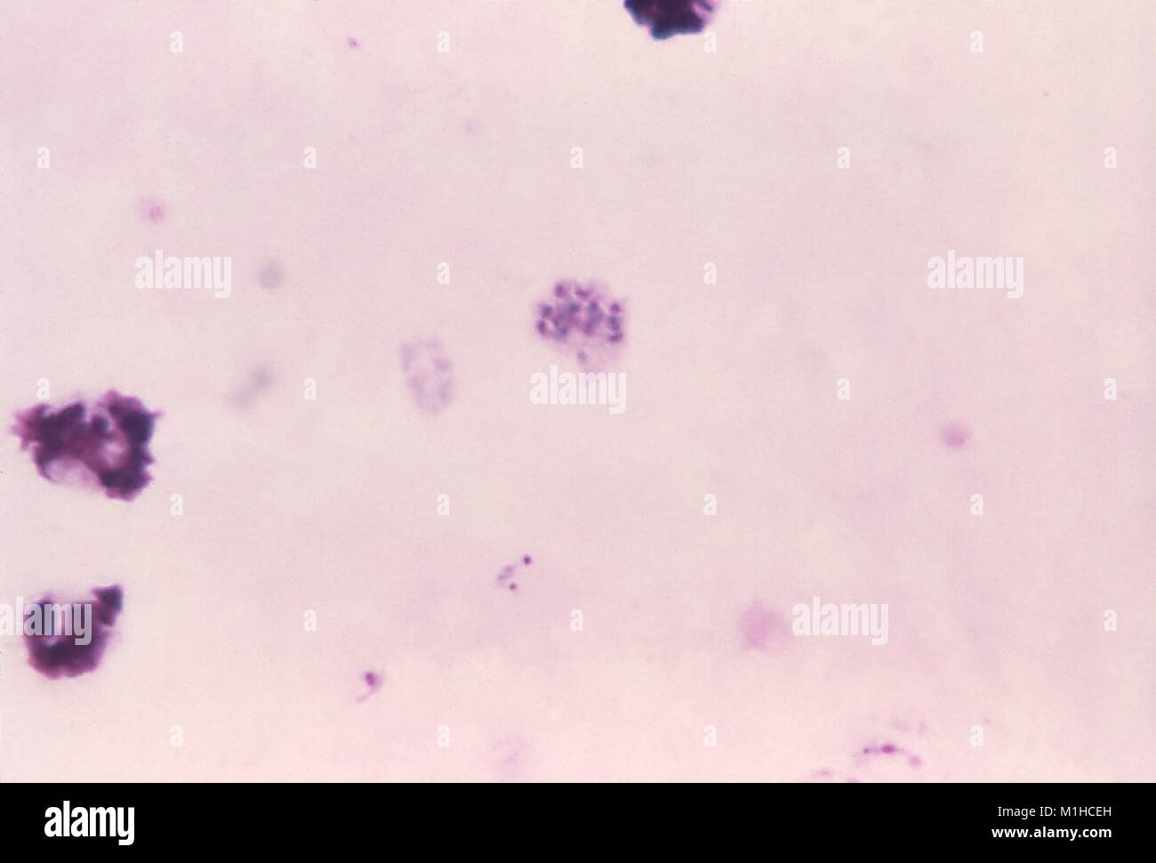
Plasmodium vivax schizont revealed in thick micrograph film using Giemsa  stain, 1971. Image courtesy Centers for Disease Control (CDC) / Dr Mae  Melvin Stock Photo - Alamy
Plasmodium vivax schizont revealed in thick micrograph film using Giemsa stain, 1971. Image courtesy Centers for Disease Control (CDC) / Dr Mae Melvin Stock Photo - Alamy

Wright-Giemsa stain | VWR